

ORIGINAL RESEARCHES
Purpose. The general goal of this methodological article, consisting of two parts, is to provide a unifying theoretical approach to the still debated problem on determining the depth of laser light penetration into biotissues and the dosage of laser therapeutic effect from the standpoint of modern medical physics. The purpose of the second part of the article is to demonstrate that calculation of the absorbed dosage at laser therapy sessions is similar to the calculation of classical doses in radiobiology and radiation therapy.
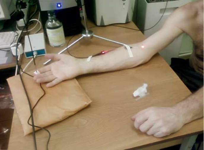
Materials and methods. The authors reviewed current state of terms and definitions related to the calculation of doses in ionizing and non-ionizing radiation. The Monte Carlo method was used to simulate the soft tissue volume in which 95 % of radiation energy is absorbed. A classical absorbed dose measured in Grays was estimated. Numerical simulation of absorbed doses for various typical laser therapy procedures was performed.
Results. It has been shown that the effective irradiated volume of tissues, despite of small variations in soft tissue density between patients, allows to calculate the absorbed radiation dose in Grays, similar to radiobiological doses. Comparative findings on a single local absorbed dose for various percutaneous therapeutic procedures do not contradict the known clinical data, and even more, make the relationship of different doses for different therapeutic purposes more clear. As it has been found, typical doses range from 0.7 Gy for intravascular blood irradiation to 106 Gy for destructive photodynamic therapy and UV therapy procedures in dermatology.
Conclusion. The proposed methodological approach proposes a new look at both the problem of the depth of laser light penetration into biotissues and the problem of laser light doses during therapeutic and diagnostic procedures from a unified medical and physical standpoint.
Objective: to compare outcomes after treatment of venous malformations (VM) in the maxillofacial region using diode laser light and sclerotherapy with 3 % Aethoxysclerol foam.
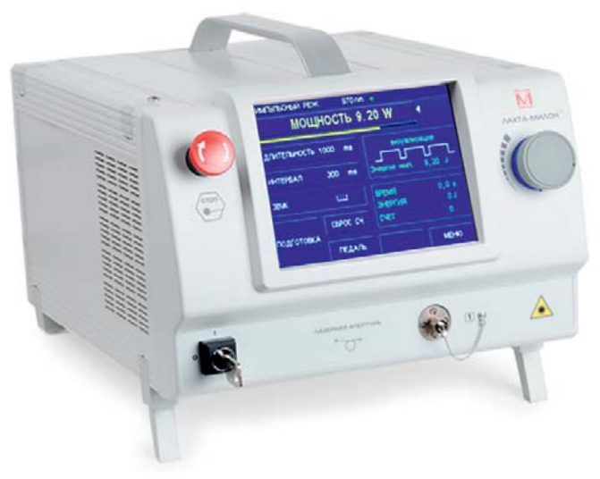
Materials and methods. 40 patients with venous malformations of the maxillofacial region were enrolled in the study. Patients were divided into two equal groups of 20 individuals each: in Group 1, patients were treated with diode laser light; in Group 2, patients had sclerotherapy with 3 % Aethoxysclerol foam. For laser therapy, Lachta-Milon diode laser (Russia) with an optical light guide having a flat end of 0.4–0.6 mm, wavelength 980 nm, pulse-periodic mode with interval 0.1–0.25 seconds at power 3.5–5.5 was used. In Group 2 VM sclerotherapy by L. Tessari’s technique (2000) in the maxillofacial region consisted of injection of microfoam made of 3 % Aethoxysclerol solution into the malformation lumen.
Results. In all patients, management was successful without intraoperative or postoperative bleeding. The performed study revealed that sclerotherapy with Aethoxysclerol foam is most effective for treating large and medium-sized malformations resulting in significant reduction in formation size. To treat malformations located in difficult anatomical areas, such as peri-orbital or hard and soft palate, diode laser with wavelength 980 nm and power ranging from 3.5 to 5.5 W turned to be the most effective.
Conclusion. Endovascular laser obliteration is an effective and safe technique for treating venous malformations in the maxillofacial region. It is also a method of choice for malformations located in the peri-orbital area.
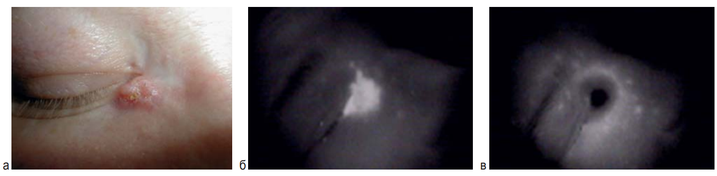
Purpose. To evaluate immediate and long-term outcomes after photodynamic therapy (PDT) of basal cell carcinoma (BCC) on the eyelid with intravenous administration of photosensitizer (PS) chlorine E6.
Materials and methods. PDT was performed in 261 patients with a verified diagnosis of BCC of the eyelid cT1-3N0MO, stage I–II, aged 38 to 87 (average 68.7 ± 15.6). PS chlorin E6 (Photolon, Photoditazin, Radachlorin) was used intravenously at dosage 1.0–1.5mg/kg; 1–2 courses. Light dosage ranged from 100 to 300 J/cm2, depending on the neoplasm clinical form and the data of fluorescence diagnostics.
Results. Complete regression after the first PDT course of eyelid BCC cT1-3N0M0 with intravenous administration of PS chlorin E6 was obtained in 90.4 % of cases; after 2 courses - in 96.2 % of cases. During the follow-up period from 5 to 15 years, relapses were diagnosed in 7.6 % of cases. The number of relapses depended on the process stage. In neoplasms corresponding to the symbol cT1a, the number of relapses was 1.4 %, cT1 b – 5.0 %, cT1 c– 8.3 %, cT2 a – 2.5 %, cT2 b – 5.0 %, cT2 c – 9 %, cT3 a – 5.8 %, cT3 b – 43.8 %, cT3 c – 46.7 %. Good and excellent cosmetic results were seen too.
Conclusion. PDT of eyelid BCC of stage I–II with intravenous administration of PS chlorine E6 is an effective and low-risk method for anatomical and functional disorders. It can be an alternativetechnique to surgical treatment. The discussed way of treatment should be personalized and provided in specialized medical settings.
REVIEW
Mechanisms of endovasal laser coagulation (EVLC) applied in varicose vein disease are not fully understood.
Purpose. To analyze currently applied EVLC mechanisms so as to prevent hemorrhagic complications and paresthesia caused by these mechanisms.
Methods. This review analyses modern theories on EVLC mechanisms when applied in varicose vein disease in the lower extremities.
Results. Published experimental and clinical trials, including histological ones, have shown that the degree of vein damage during EVLC session depends on many factors, such as wavelength, intensity, and optical fiber speed. Damage to veins during EVLC procedure depends on various factors, such as direct contact of the vein wall with an optical fiber tip, carbonization of blood elements leading to the increased intravenous blood temperature and to the formation of gas bubbles as well as heat convection on the vein wall through the blood.
Conclusion. Destruction of the vein wall during EVLC procedure is the result of a synergistic effect of various damaging factors. Currently, 2-μm laser irradiation is being implemented into clinical practice. This technique provides better vein coagulation under less power values which promotes less postoperative complications.
A CLINICAL OBSERVATION
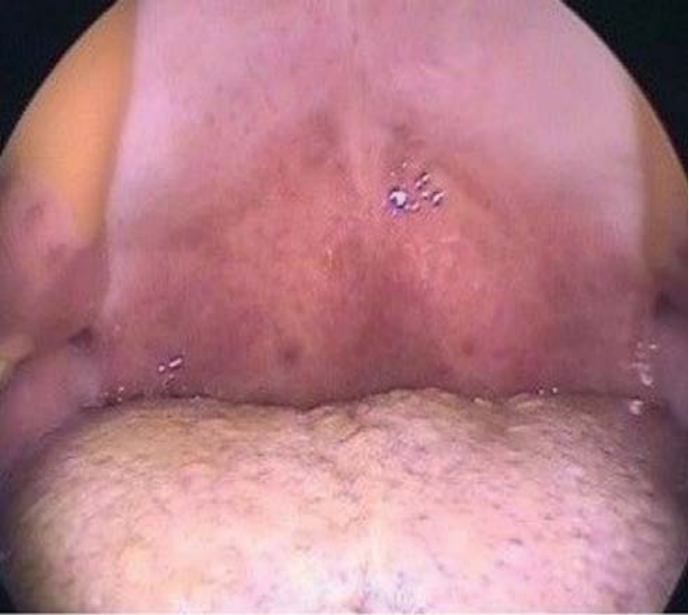
Objective. To present a clinical case of successful application of laser sculptural uvulopalatoplasty in a patient with ronchopathy and severe obstructive sleep apnea syndrome.
Materias and methods. The article describes a clinical case of successful application of soft palate surgery – laser sculptural uvulopalatoplasty – in a patient with ronchopathy and severe obstructive sleep apnea syndrome.
Results. After the described surgical intervention, rehabilitation stage was wihtout complications. A complete relief from night snoring and apnea in the patient has been achieved. The obtained results and the absence of complications after surgery were registered in the patient for the next 5 years and more.
Conclusion. Laser sculptural uvulopalatoplasty is a safe and effective method of treating patients with ronchopathy and obstructive sleep apnea syndrome, even severe one, which provides a stable and complete relief from night snoring and its complications.
On the 70th anniversary of his birth
OBITUARY

ISSN 2686-8644 (Online)


