

ORIGINAL RESEARCHES
Purpose. The general purpose of present methodological article, consisting of two parts, is to provide a unifying theoretical approach to the still debated problem of determining the depth of penetration of laser light into tissues and the dosage of laser therapeutic effects from the standpoint of modern medical physics. The purpose of the first part of the article is to discuss the depth of laser light penetration into tissues and organs during diagnostic and therapeutic procedures and to formulate practical recommendations for its measurement.
Material and methods. The review is devoted to the current problem on the depth of laser light penetration, with terms and definitions. Based on different approximations of the radiation transfer equation known in physics, numerical theoretical estimates of the penetration depth of laser light with different wavelengths into the skin are given for different approaches. The Monte Carlo method was used to simulate soft tissue volume in which radiation energy is absorbed up to 95 %. The depth of light penetration was estimated using linear dimensions of the volume.
Results. As it turned out, the classical theoretical depth of laser light penetration into tissues and organs highly depends not only on wavelength and tissue optical properties, but also on the chosen approximation and calculation method. The penetration depth, defined by the calculated volume in which up to 95 % of laser radiation is absorbed, is about 3 times greater than the classical theoretical penetration depth, which better complies with known experimental findings.
Conclusion. The depth of laser light penetration into tissues can be more reasonably determined for therapeutic and diagnostic procedures via the effective irradiated volume of tissues.
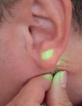
Background. Currently, the problem of radical removal of pigmented skin formations with good clinical and aesthetic results remains open. These formations, including congenital giant pigmented nevi (CGPN), are found at birth in 2 % of newborns; acquired nevi are registered in 75 % of children. They can be large in size and quite often are located on visible parts of the body, creating psychological problems in children and leading to aesthetic discomfort. In adulthood, various complications may develop including malignancy which may develop in 10% of patients. Management of pigmented skin formations causes both medical and psychological problems. So, it is reasonable to solve them in childhood. Modern various techniques for the removal of pigmented nevi are not always effective; besides, they are often followed by various complications, like relapses (in 6–41 %) and skin scarring (in 6 %). Such a situation requires development of new highly effective techniques for removing various pigmented nevi, including laser light. Recently, separate works on the application of “blue” (λ = 450 nm) laser light and infrared (λ = 10.6 µm) laser light generated by periodic CO2 laser for the discussed pathology have appeared.
Purpose. To conduct a comparative experimental study on the effects of “blue” (λ = 450 nm) laser light and infrared (λ = 10.6 µm) laser light generated by pulsed periodic CO2 laser at the skin of laboratory rats in order to find out promising parameters for surgical removal of pigmented skin formations.
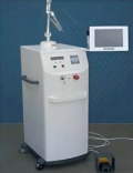
Material and methods. Laser devices “Lasermed 10-03” (LLC RIK, Russia) generating in “blue” (λ = 450 nm) and pulsed periodic CO2 laser “ALDAN” (IOF RAS, Russia) generating in infrared (λ = 10.6 µm) laser light were used. In present in vivo experiment, pigmented skin of laboratory rats was exposed to laser light. Exposure zones were compared; features and regeneration terms were analyzed too.
Results. The researchers registered specific effects in skin irradiation with laser light as well as in terms of wound regeneration. They also identified optimal parameters of “blue” (λ = 450 nm) and infrared (λ = 10.6 µm) laser light for the removal of CGPN and pigmented skin formations.
Conclusion. The obtained results of the present comparative experimental trial have outlined prospects for the application of “blue” (λ = 450 nm) laser light and infrared laser light (λ = 10.6 µm) generated by pulsed periodic CO2 laser for surgical removal of pigmented skin formations.
Purpose: to background the reasonability in performing laser sculptural uvulopalatoplasty in patients with ronchopathy and obstructive apnea sleep syndrome.
Materials and methods: the authors back grounded the application of laser sculptural uvulopalatoplasty in patients with ronchopathy and obstructive apnea sleep syndrome. This technique allows to minimize surgical trauma of the tendon-muscle plate during vaporization of pathologically altered soft palate tissues which is confirmed by outcomes after surgery in 309 patients with ronchopathy and obstructive sleep apnea syndrome who were under follow-up observation for 5 years and more.
Results: outcomes after laser sculptural uvulopalatoplasty demonstrate not only complete relief of patients of ronchopathy and obstructive sleep apnea syndrome (result of night snoring) and related complications, but also demonstrate safety and high efficiency (98.4%) of this surgical intervention and stable preservation of the obtained results during 5 years or more after surgery.
Conclusion: laser sculptural uvulopalatoplasty, performed under the careful observation of surgical technique and after careful selection of patients after comprehensive differentiated examination is a safe and effective method for treating patients with ronchopathy and obstructive sleep apnea syndrome. Thus, patients can have stable and complete relief of night snoring and its complications.
CURRENT INFORMATION

ISSN 2686-8644 (Online)


